Clinical trial: intravenous pantoprazole vs. ranitidine for the prevention of peptic ulcer reb-leeding: a multicentre, multinational, randomized trial
Статьи Опубликовано в журнале:«Alimentary Pharmacology & Therapeutics»; 29; 497-507.
C. Van Rensburg1, A. N. Barkun2, I. Racz3, R. Fedorak4, P. C. Bornman5, C. Begli Nger6, J. Balanzo7, J. Deviere8, L. Kupcinskas9, R. Luehmann10, H. Doerfler10 S. Schafer-preuss10
2Department of Medicine, Division of Gastroenterology, McGill University, Montreal, Quebec, Canada;
3Petz Aladar Teaching and County Hospital, Gyor, Hungary;
4University of Alberta, Edmonton, Alberta, Canada;
5Grootte Schuur Hospital, Cape Town, South Africa;
6University Hospital Basel, Basel, Switzerland;
7Hospital Santa Creu Sant Pau, Barcelona, Spain;
8U.L.B. Hospital Erasme, Brussels, Belgium;
9Kaunas University of Medicine, Kaunas, Lithuania;
10Nycomed GmbH, Konstanz, Germany
SUMMARY
Background
Controlled pantoprazole data in peptic ulcer bleeding are few.
Aim
To compare intravenous (IV) pantoprazole with IV ranitidine for bleeding ulcers.
Methods
After endoscopic haemostasis, 1256 patients were randomized to pantoprazole 80 mg+8 mg/h or ranitidine 50 mg+13mg/h, both for 72 h. Patients underwent second-look endoscopy on day 3 or earlier, if clinically indicated. The primary endpoint was an overall outcome ordinal score: no reb-leeding, reb-leeding without/with subsequent haemostasis, surgery and mortality. The latter three events were also assessed separately and together.
Results
There were no between-group differences in overall outcome scores (pantoprazole vs. ranitidine: S0: 91.2 vs. 89.3%, S1: 1.5 vs. 2.5%, S2: 5.4 vs. 5.7%, S3: 1.7 vs. 2.1%, S4: 0.19 vs. 0.38%, P = 0.083), 72-h clinically detected reb-leeding (2.9% [95% CI 1.7, 4.6] vs. 3.2% [95% CI 2.0, 4.9]), surgery (1.9% [95% CI 1.0, 3.4] vs. 2.1% [95% CI 1.1, 3.5]) or day-3 mortality (0.2% [95% CI 0, 0.09] vs. 0.3% [95% CI 0, 1.1]). Pantoprazole significantly decreased cumulative frequencies of events comprising the ordinal score in spurting lesions (13.9% [95% CI 6.6, 24.7] vs. 33.9% [95% CI 22.1, 47.4]; P = 0.01) and gastric ulcers (6.7% [95% CI 4, 10.4] vs. 14.3% [95% CI 10.3, 19.2], P = 0.006).
Conclusions
Outcomes amongst pantoprazole and ranitidine-treated patients were similar; pantoprazole provided benefits in patients with arterial spurting and gastric ulcers.
INTRODUCTION
Peptic ulcer disease accounts for 50-70% of acute nonvariceal upper gastrointestinal (GI) bleeding.1 Early endoscopy and haemostasis, where appropriate, have proven beneficial for patients with bleeding peptic ulcers at both low and high risks.1-3 However, an estimated 20% of patients will have persistent or reb-leeding following endoscopy and up to 10% will require surgery. 4,5
Although H2-receptor antagonists (H2RAs) have shown modest improvement in reb-leeding6, 7 restricted to patients with gastric ulcers, inadequate acid suppression and tolerance limit the benefit of these agents.8, 9 Meta-analyses suggest that the superior acid suppression seen with proton pump inhibitors (PPIs) translates into lower rates of reb-leeding and surgery compared with placebo or H2RAs, although there exists heterogeneity between the different trials and controversy and uncertainty as to optimal dosing.5, 10, 11 As a result, high-dose IV PPI is now recommended in the management of patients with nonvariceal upper GI bleeding.3 However, fully published double-blind, randomized trials using IV pantoprazole, the most commonly used IV PPI in North America, remain few and two trials suggested a benefit of a high-dose PPI over H2RAs12, 13 while the other did not.14 The current study was thus designed to investigate the safety and efficacy of high-dose intravenous pantoprazole15 compared with high-dose intravenous ranitidine16 for the prevention of acute peptic ulcer reb-leeding following successful endoscopic haemostasis in-patients with actively bleeding lesions or nonbleeding visible vessels.
MATERIALS AND METHODS
Study design and objectives
This study was conducted as a multicentre, randomized, controlled, double-blind, parallel-group comparison across 137 centres in 15 countries between March 2000 and October 2001. Standardized protocol procedures were adopted by all participating centres. Patients who underwent successful endoscopic haemostasis of bleeding peptic ulcers were considered for inclusion in the trial. The participants were enrolled by the individual sites after a review of selection criteria. A blinded random allocation was carried out centrally according to computer-generated random block numbers.
The objective of the study was to compare the efficacy and safety of intravenous pantoprazole and ranitidine in-patients with bleeding peptic ulcers after successful endoscopic haemostasis.
Selection criteria
Hospitalized adults aged 18 years or older who underwent successful endoscopic haemostasis for a bleeding gastric or duodenal peptic ulcer were eligible for entry if active spurting (Forrest Ia), oozing (Forrest Ib), or a non-bleeding visible vessel (NBVV, Forrest IIa)17 was noted at endoscopy. Because of the controversy surrounding endoscopic treatment of adherent clots,3 patients presenting with a clot in the ulcer base could only be entered into the study if, when removed, the underlying ulcer lesion was reclassified as Forrest Ia, Ib or IIa.17 If the clot could not be removed, the patient was deemed ineligible. The various ethics committees reviewed and approved the protocol and all patients were required to give written informed consent.
Patients were excluded, if they had oesophageal varices, portal hypertension, Child's C liver cirrhosis or concomitant disease that made inclusion inappropriate (e.g. terminal disease, malignancy of GI tract, GI bleeding from other sources). The following medications that might have a confounding effect on reb-leeding were not permitted: H2RAs, PPIs, somatostatin, misoprostol, sucralfate, prokinetics or antacids from the time of the patient's admission until the end of study treatment. Other exclusion criteria were the need for anticoagulants during the study period, use of concomitant medications with the potential for drug interactions, pregnancy, lactation, child-bearing potential not using adequate contraception or allergy to ranitidine or pantoprazole. Helicobacter pylori positive patients were also included in the study, but eradication was not permitted during the 72-h treatment period, as this might have affected ulcer healing and acted as a confounder of outcome.18
Endoscopic treatment
In an attempt to optimize endoscopic haemostasis and allow for broadly generalizable results, acceptable methods of endoscopic haemostasis included epinephrine:saline 1:10 000 with or without sclerosant injection, electro- or thermocoagulation (bipolar electrocoagulation or heater probe), fibrin glue, or epinephrine:saline 1:10 000 injection combined with one of these methods (the study was carried out before the diffusion of endoscopic clips). Rather than enforcing a specific endoscopic haemostatic method on participating endoscopists, this pragmatic approach allowed for an acceptable method of haemostasis they were familiar with. By allowing this, the study results also gained generalizability.
If endoscopic haemostasis was judged to be successful, eligible patients were randomized to either pantoprazole 80 mg bolus followed by 8 mg/h continuous infusion or ranitidine 50 mg bolus followed by 13 mg/h continuous infusion both for 3 days. The double-blind design was achieved by using pantoprazole-placebo lyophilisates or ranitidine-placebo solutions for patients assigned to ranitidine or pantoprazole treatment arms respectively, in addition to the active drug preparations appropriate to the treatment allocation. An in-line filter was not a requirement for the formulation of intravenous pantoprazole that was used. Transfusion practices, a decision on the need for surgery and the subsequent treatment after the 72-h intervention were left at the discretion of the investigator according to standard practice for the institution and in keeping with the medical effectiveness philosophy of the trial to increase the generalizability of results.
Assessment of outcomes
Clinical suspicion of reb-leeding was defined as any one of the following three signs: (i) vomiting of fresh blood, (ii) insufficient increase in haemoglobin or increase in need for blood transfusions, or (iii) haemodynamic instability (two of the following: decrease in haemoglobin to <10 g/dL or a drop ≥2 g/dL, increase in heart rate >100 beats/min or of >20 beats/min from baseline or decrease in systolic blood pressure <100 mm Hg or of a drop >20 mm Hg from baseline) similar to criteria adopted in previous trials.5 Patients exhibiting signs of reb-leeding then underwent either an emergency endoscopic procedure or surgery using an effectiveness-trial approach (i.e. according to local existing practice) to assess further the possibility of reb-leeding (except for the presence of haematemesis in isolation); if not, a routine 'second-look' endoscopy was performed at 72 h. Clinical suspicion of reb-leeding (as previously defined) within 4 h after the initial attempt at endoscopic haemostasis was not classified as reb-leeding, but rather as failure of endoscopic therapy. The prespecified primary and secondary outcomes were based on an overall composite outcome score. In addition, the cumulative frequencies of clinically suspected reb-leeding confirmed endoscopically, surgery and mortality were analysed and performed. A post hoc analysis was performed to allow comparisons with other trials; to do so, it adopted more the commonly used outcomes of clinically suspected reb-leeding confirmed at endoscopy, surgery and mortality.
Primary outcome measure. Because of the performance of routine second-look endoscopy, we could not adopt the traditional outcome of clinically suspected reb-leeding as primary outcome. Thus, the primary efficacy measure chosen was the ordinal overall outcome score (see below) as assessed during an earlier unscheduled or at the scheduled routine second-look endoscopy at 72 h. A similar overall outcome score as was used in other published trials19'20 was scored from best to worst using the following ordinal ranking scale: 0 = no reb-leeding; 1 - minor reb-leeding not requiring endoscopic haemostasis; 2 - major reb-leeding requiring additional endoscopic haemostasis; 3 - reb-leeding requiring surgery; 4 - reb-leeding causing death.
Secondary outcome measures. The secondary outcome measures included the proportion of patients with each of the possible outcome events (scores 1, 2, 3, or 4) in each group, the number of blood units transfused after randomization and mortality at 14 days (both overall and attributable to reb-leeding as determined by a blinded data review committee).
A priori defined subgroup analysis looked at the cumulative frequencies of the four outcome events comprising the overall outcome score, while adjusting for Forrest classification (Ia, Ib, IIa), site of the bleeding ulcer (gastric, duodenal), H. pylori status, age, ulcer history, method of endoscopic haemostasis and the continuous use at study entry of nonsteroidal anti-inflammatory drugs (NSAIDs) (continuous intake), low-dose aspirin (<100 mg/day) or anticoagulants.
Post hoc analysis. The results were also analysed a posteriori according to the more commonly used outcomes of clinically suspected reb-leeding and surgery or mortality (due to reb-leeding at 72 h). For this analysis, clinically suspected reb-leeding was defined using only the clinical suspicion of reb-leeding that subsequently required endoscopic retreatment, as previously described in the literature.3 Rebleeding detected only at the 72-h planned endoscopy that required endoscopic treatment was not included in this post hoc analysis for the specific purpose of comparing results with other published trials.
Additional symptoms and adverse events. General gastrointestinal symptoms were assessed at baseline, day 1 and day 3 and included heartburn, acid regurgitation, dysphagia, epigastric pain, abdominal pain, burping/belching, nausea, retching and vomiting. Additional secondary outcomes included adverse events, as well as laboratory test, heart rate and blood pressure measurements. Adverse events were listed and the investigator's causality assessments reported; in addition, the sponsor's regulatory documentation in this regard was also analysed seeking all adverse events.
Sample size calculations and planned statistical analyses
It was planned to enroll 1200 patients to obtain 1000 per protocol patients for evaluation. A sample size of 500 evaluable patients in each group has 95% power to detect a probability of 0.566 that an observation in one group was less than an observation in the other group, using a Wilcoxon (Mann-Whitney) rank-sum test with a 0.050 two-sided significance level. Testing for non-inferiority was also planed as initial step with a non-inferiority margin set at 0.45 of the relative effect. Considering possible drop-outs, a total of 628 patients were to be enrolled in each group.
Standard descriptive statistics were applied to safety variables including adverse events, laboratory tests, heart rate and blood pressure. All between-groups comparisons were analysed using Fisher's Exact test for categorical variables and Mann-Whitney U-test for ordinal variables.
All analyses were planned a priori with the exception of the post hoc analysis as previously defined. Outcome events (scores 1-4) were compared using the Cochran-Mantel/Haenszel methods. Stratified analyses for this outcome took into consideration individual covariates only if at least 30 patients were reached per stratum and treatment group. Between-group comparisons in outcome events were stratified by Forrest classification (Ia, Ib, IIa), site of the bleeding ulcer (gastric, duodenal), H. pylori status, age, ulcer history, method of endoscopic haemostasis and premedication with nonsteroidal anti-inflammatory drugs (NSAIDs) (continuous intake), low-dose aspirin (<100 mg/day) or anticoagulants.
All-cause mortality and mortality attributable to bleeding were analysed by survival analysis using the Kaplan-Meier Life-Table method, both at days 3 and 14 following inclusion in the trial. The 95% confidence intervals for the between-group differences were determined.
The per-protocol (PP) population included all patients without a major protocol violation during the study, whereas the intention-to-treat analysis (ITT) and modified ITT populations included all patients with bleeding peptic ulcers who underwent successful endoscopy and received at least one dose of study drug, including patients with incomplete data. In the ITT analysis, patients with missing follow-up endoscopy data were imputed the 'worst case' score of 4; this was not done in the modified ITT analysis. In keeping with the original protocol specifications, the primary endpoint (overall outcome ordinal score) is reported for the ITT population. The post hoc analysis in contrast is reported as per the modified ITT approach as it would otherwise be meaningless (with an attribution of death to all patients with missing data) and is indeed in keeping with other trials to allow for comparison with published literature. All conclusions were similar using PP, ITT or modified ITT analyses.
The safety set (used in analyses of mortality and safety) comprised of all patients receiving at least one dose of study medication including patients who did not meet the primary entry criterion of a bleeding peptic ulcer. Safety data are reported on the safety set, in keeping with regulatory requirements. The statistical package used was SAS version 8.2 (SAS Institute, Cary, NC, USA).
RESULTS
Patient description
Between March 2000 and October 2001, the 137 centres screened 3255 patients of whom 1999 did not meet study entry criteria. Participating countries included: (Austria, Belgium, Canada, France, Germany, Great Britain, Hungary, Italy, Lithuania, Netherlands, Poland, Portugal, Spain, South Africa and Switzerland). The most common reasons for screening failure were the presence of Forrest IIb, IIc, III (clot that could not be removed, a flat spot or no stigmata respectively) lesions (22.4%), upper GI varices as cause of bleeding (15.1%), multiple GI findings (13.4%) and GI bleeding attributable to non-ulcer sources (10.4%). As a result, 1256 patients were randomized to receive at least one dose of study medication and were included in the safety analysis. Of these, 12 patients did not meet the primary entry criterion, leaving 1244 for the ITT analysis. An additional cohort of 186 patients were excluded because of protocol violations, the most frequent of which were medication errors, disallowed previous/concomitant medications and delayed or no final endoscopy. Overall, only 48 patients had protocol deviations that led to study discontinuation (16 received pantoprazole and 32 ranitidine). Follow-up endoscopy was not performed in 12 and 21 patients in the pantoprazole and ranitidine groups respectively. The full CONSORT diagram is included in Figure 1.
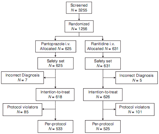
Figure 1. CONSORT diagram.
There were no statistically significant differences between the two treatment groups in terms of demographic and pre-treatment clinical characteristics (Table 1). At index endoscopy, 65% of patients had active bleeding (spurting: 10%, oozing: 55%) and 35% had nonbleeding visible vessels. The most common method of haemostatic treatment was epinephrine:saline 1:10 000 injection alone, which was performed in 64% of cases; 23% of patients received a combination of two or more haemostatic treatments.
Efficacy
Primary outcome. Pantoprazole showed a numerically lower (better) overall outcome score at day 3 compared with ranitidine (in 612 and 633 patients, respectively), but the difference did not achieve statistical significance (scores for pantoprazole and ranitidine respectively were S0: 91.2 vs. 89.3%, S1: 1.5 vs. 2.5%, S2: 5.4 vs. 5.7%, S3: 1.7 vs. 2.1%, S4: 0.19 vs. 0.38, P = 0.083). Pantoprazole did fulfil the preset criteria of non-inferiority compared to ranitidine.
Secondary outcomes. There were no differences in 72-h clinically detected surgical rates (1.9% [95% CI 1, 3.4] vs. 2.1% [95% CI 1.1, 3.5]). The 3 and 14-day all-cause mortality rates did not differ significantly between the pantoprazole and ranitidine groups being 0.2% (95% CI 0, 0.9) vs. 0.3% (95% CI 0, 1.1) and 1.5% (95% CI 0.5, 2.4) vs. 2.6% (95% CI 1.3, 3.8) respectively. The odds ratio for the comparison of 3-day all-cause mortality was 0.34 (95% CI 0.04, 3.6). Similarly, the day-3 and day-14 mortality rates attributable to reb-leeding were not different: 0.2% (95% CI 0.0, 0.5) vs. 0.5% (95% CI 0, 1.0) and 0.5% (95% CI 0, 1.0) vs. 0.6% (95% CI 0, 1.3) in the pantoprazole vs. ranitidine group respectively.
Table 1.
Baseline characteristics
| Clinical characteristic | Pantoprazole (n = 618) | Ranitidine (n = 626) |
| Female (%) | 32 | 30 |
| Age, median (y) (Min-Max) | 63 (18-95) | 63 (18-97) |
| ≥60 years (%) | 57.4 | 56.9 |
| Caucasian (%) | 89.6 | 90.5 |
| Asian (%) | 1.4 | 2.1 |
| BMI, median (kg/m2) | 25.3 | 25.4 |
| Smokers (%) | 26.3 | 26.6 |
| Alcohol (%) | 13.9 | 16.0 |
| Cardiovascular disease (%) | 3.5 | 4.8 |
| H. pylori positive (%) | 79.1 | 77.5 |
| Previous peptic ulcer bleed (%) | 16.3 | 18.7 |
| First occurrence of peptic ulcer (%) | 59.9 | 60.4 |
| Relapse occurrence of peptic ulcer (%) | 40.1 | 39.6 |
| Prior NSAIDs (%) | 29.5 | 31.5 |
| Prior low-dose aspirin (%) | 11.2 | 8.6 |
| Prior anticoagulants (%) | 28.5 | 31.8 |
| Hemodynamic instability (%) (BP: systolic ≤100 or diastolic <60 mm Hg) | 12 | 12 |
| Site of ulcer | ||
| Gastric (%) | 43.4 | 41.4 |
| Duodenal (%) | 56.1 | 58.3 |
| Forrest distribution | ||
| Ia (spurting) (%) | 10.5 | 9.4 |
| Ib (oozing) (%) | 53.4 | 55.9 |
| IIa (NBVV) (%) | 36.1 | 34.7 |
There was no significant difference in transfusion requirements between the two groups (54% of patients in the pantoprazole and 50% in the ranitidine group were transfused requiring a median of 2 units in each group with 7.6% and 8.5% respectively requiring 5 units or more P = 0.18).
No patient experienced bleeding within the first 4 h following endoscopy.
Planned analysis according to the cumulative frequency of outcome events that comprise the overall score (S1-4) showed that Forrest staging and the site of peptic ulcer bleeding at initial endoscopy were found to influence outcome events significantly. For patients diagnosed with initial Forrest Ia, peptic ulcer bleeding (active spurting bleeding, total n = 124), treatment with pantoprazole (13.9%; 95% CI 6.6, 24.7) led to significantly fewer outcome events compared with patients treated with ranitidine (33.9%; 95% CI 22.1, 47.4; P =0.01). Pantoprazole also significantly reduced the incidence of outcome events among patients with gastric (total n = 527, 6.7%; 95% CI 4.0, 10.4 vs. 14.3%; 95% CI 10.3, 19.2; P = 0.006), but not duodenal ulcers (total n =712, 14.4% vs. 14.3%) compared with ranitidine. No other comparisons of treatment groups were significant with regard to the effect of H. pylori status, age, ulcer history, method of endoscopic haemostasis, premedication with NSAIDs, low-dose aspirin (<100 mg/day) or anticoagulants.
Rebleeding was noted in identical numbers of pantoprazole and ranitidine-treated patients when assessing the primary outcome according to Forrest Ib (22% in each group) and Forrest IIa (19% in each group) classification.
Post hoc analyses of traditional outcomes. There were no statistically significant between-group differences in clinically suspected reb-leeding requiring haemostasis, surgery or mortality due to reb-leeding (Table 2). Clinically suspected reb-leeding requiring retreatment on endoscopy occurred in 2.9% (95% CI 1.7, 4.6) of patients in the pantoprazole group and 3.2% (95% CI 2.0, 4.9) in the ranitidine group. Of note, reb-leeding requiring endoscopic treatment that was detected only at the follow-up endoscopy occurred in 2.4% (95% CI 1.4, 4.0) in the pantoprazole and 2.9% (95% CI 1.7, 4.5) in the ranitidine patients. Therefore, the total rate of reb-leeding requiring endoscopic retreatment was 5.3% (95% CI 3.7, 7.4) and 6.1% (95% CI 4.3, 8.2) for the pantoprazole and ranitidine groups respectively (Table 2).
Overall, 92% of subjects in the ITT population had the pre-planned 72-h endoscopy and of these 2.9% (95% CI 1.9, 3.9) required additional endoscopic haemostatic therapy with no differences between treatment groups. This accounted for 46.5% (95% CI 34.5, 58.7) of the reb-leeding requiring retreatment.
Attributable surgery (1.9% [95% CI 1.0, 3.4] vs. 2.1% [95% CI 1.1, 3.5]) and 3-day mortality (0.2% [95% CI 0.0, 0.9] vs. 0.3% [95% CI 0.0, 1.1]) did not differ between pantoprazole and ranitidine groups respectively (Table 2).
Analyses of traditional outcomes according to initial Forrest class did not reveal any statistically significant difference between the groups. However, more than twice as many patients with Forrest Ia (spurting) stigmata developed clinically suspected reb-leeding in the ranitidine group compared with the pantoprazole group (11.9%; 95% CI 4.9, 22.9 and 4.6%; 95% CI 0.96, 12.9; P = 0.25). When analysing results according to ulcer location, no patient with gastric ulcer developed clinically suspected reb-leeding in the pantoprazole group (0%; 95% CI 0.0, 2.1), compared to 10 (3.9%; 95% CI 1.9, 7.0; P = 0.01) patients in the ranitidine group. In-patients with duodenal ulcers, no statistical significant difference between the treatment groups was seen pantoprazole 5.2%; (95% CI 3.1, 8.1) vs. ranitidine 2.7% (95% CI 1.3, 5.0) (p = 0.14).
Medication-related side effects
Similar proportions of patients (42% for pantoprazole vs. 44% for ranitidine) reported adverse events (Table 3). The most common events were headache, insomnia, phlebitis, hypertension, constipation and anxiety, of which most were assessed as unrelated to the study medication. Significantly more patients in the ranitidine group (n = 19, 1.1%; 95% CI 0.5, 2.3) than in the pantoprazole group (n = 7, 3.0%; 95% CI 1.8, 4.7) discontinued the study prematurely due to adverse events; however, the investigator assessed most cases as 'unrelated' to the study medication. There were more adverse events rated as 'likely' or 'definitely' related to the study medication in the pantoprazole group (6.5% vs. 2%; P = 0.0035). This was due in large part to the higher incidence of injection site reactions in the pantoprazole group, which accounted for 13 of the 27 treatment-related adverse events. Overall, regardless of causality, injection site reactions (primarily mild to moderate superficial thrombophlebitis) occurred in 5.3% of patients in the pantoprazole group and 0.5% in the ranitidine group (p < 0.0001). Only one case was rated as severe. The proportion of patients with serious adverse events was similar in both treatment groups (pantoprazole 3.2%; 95% CI 2.0, 4.9 and ranitidine 4.8%; 95% CI 3.2, 6.7) (Table 3).
Table 2.
Outcomes of bleeding peptic ulcers after 72 h of treatment in the modified ITT population (post-hoc analysis)
| Pantoprazole (n = 618) (% [95% CI]) | Ranitidine (n = 626) (% [95% CI]) | P-value | |
| Clinically suspected reb-leeding (detected via clinical suspicion of reb-leeding) that required treatment | 18 (2.9 [1.7, 4.6]) | 20 (3.2 [2.0, 4.9]) | 0.90 |
| Surgery due to reb-leeding | 12 (1.9 [1.0, 3.4]) | 13* (2.1 [1.1, 3.5]) | 0.97 |
| Mortality due to reb-leeding (Day 3) | 1 (0.2 [0.0, 0.9]) | 2 (0.3 [0.0, 1.1]) | 0.99 |
| Total haemostatic retreatment** | 33 (5.3 [3.7, 7.4]) | 38 (6.1 [4.3, 8.2]) | 0.87 |
** Rebleeding requiring retreatment detected solely at the time of the scheduled second-look endoscopy accounts for the remaining patients having undergone endoscopic haemostatic retreatment. They totaled 2.4% (95% CI 1.4, 4.0) in the pantoprazole and 2.9% (95% CI 1.7, 4.5) in the ranitidine group (P = 0.75).
Table 3.
Summary of adverse events
| Parameter | Pantoprazole | Ranitidine |
| (n = 625) | (n = 631) | |
| Summary of all adverse events | ||
| Number of patients with adverse events | 265 (42%) | 278 (44%) |
| Number of adverse events | 415 | 418 |
| Unrelated (Not related) | 272 (66%) | 308 (74%) |
| Unlikely related | 116 (28%) | 101 (24%) |
| Likely related | 26 (6%)* | 9 (2%) |
| Definitely related | 1 (0.2%)f | 0 |
| Summary of serious adverse events | ||
| Number of patients with SAE | 20/625(3.2%) | 30/631 (4.8%) |
| Number of SAEs | 30 | 40 |
| Unrelated (Not related) | 24 | 32 |
| Unlikely related | 5 | 8 |
| Likely related | 1** | 0 |
| Definitely related | 0 | 0 |
* Includes 13 superficial phlebitis.
**One superficial phlebitis.
The population analysed for the adverse events is the safety set as defined in the methods section.
Five (0.8%; 95% CI 0.3, 1.8) myocardial infarctions (MI) and 4 (0.6%; 95% CI 0.2, 1.6) cerebrovascular accidents (CVA) occurred among patients in the ranitidine group, while no MIs and one CVA (0.16%; 95% CI 0, 0.9) occurred in the pantoprazole group; these events were determined by the investigators to be unrelated or unlikely related to study medication.
DISCUSSION
In this study, the group of patients treated with pantoprazole exhibited a numerically lower overall outcome score (the planned primary outcome of the study) compared with those who received ranitidine. The demonstrated numerical superiority of pantoprazole over ranitidine did not reach statistical significance, even though it did fulfil criteria for non-inferiority. Post hoc analysis of traditional outcomes including clinically suspected reb-leeding and surgery or mortality due to reb-leeding also demonstrated no significant differences. The strengths of this study include use of pantoprazole in acute peptic ulcer bleeding, which has not been widely reported in the literature and the number of patients included. Indeed, this trial is the largest IV PPI trial reported to date. Additionally, only patients with high-risk endoscopic stigmata were included and all patients received endoscopic haemostasis as per current recommendations.3
A recent meta-analysis that includes the preliminary data from the current trial has suggested improved outcomes, including all-cause mortality, attributable to high-dose IV PPI in-patients at high risk of reb-leeding following endoscopic therapy.5 This trial reported 3-day outcome results as well as 14-day all-cause and reb-leeding-related mortality. The 3-day reb-leeding rate is relevant as studies on the natural history of stigmata following endoscopic therapy21 and continuous IV PPI trials12,22 have shown that the majority (57-83%) of reb-leeding occurs within the first 3 days; similarly, Jensen et al. recently reported that 56% of reb-leeding occurred in the first 72 h.14 The Cochrane meta-analysis also assessed all dosages of PPI for ulcer reb-leeding prophylaxis and reported pooled reb-leeding rates at 3 days of 8.3% for PPI treatment and 14.2% for controls.5 This result remained statistically significant in favour of PPI treatment when, by sensitivity analysis, any one of the included studies was removed; however, there was significant heterogeneity in the analysis. Focusing on trials that used high-dose intravenous bolus and continuous infusion PPI therapy, three-day reb-leeding rates have been 3-4% with PPI therapy compared to 8-16% with H2RA therapy.12, 14, 22, 23 The rate of clinically significant reb-leeding with pantoprazole (2.9%) in this study was comparable to that reported in other studies, but the rate with ranitidine (3.2%) was lower than that expected based on available literature. Although recently, Hsu et al. reported a lower overall 8-week reb-leeding rate with pantoprazole compared with ranitidine (4% vs. 16%, P = 0.04),24 Jensen et al., found no significant difference in 3-day or 30-day reb-leeding rates comparing pantoprazole IV with ranitidine IV (4.2% vs. 6.9% and 6.9% vs. 14.3% respectively).14 However, this study was terminated prematurely after enrolment of only 149 patients, and greater patient enrolment might have yielded significant differences.
Differences in findings of trials 5, 10, 11 may be related to their inclusion of Asian patients who are known to have different physiological (gastric parietal cell masses), pharmacokinetic profiles (proportions of slow vs. fast PPI metabolizers) and higher H. pylori infection rates (known to favour PPI-induced acid suppression).25 Additionally, patients in the current study may have generally been at lower risk of reb-leeding (and mortality). Indeed, only 4% of patients had a history of comorbid cardiovascular disease and only 40% had a history of previous ulcer bleeding. In fact, the rates of reb-leeding requiring haemostatic retreatment in this trial were only 5.3% with pantoprazole and 6.1% with ranitidine compared to 11% with PPI therapy and 17% with control therapies (H2RA or placebo) in the meta-analysis by Leontiadis et al.5 This lower risk of reb-leeding may be attributable to the increased initial use of modern endoscopic haemostatic treatments as nearly a quarter of patients received combination therapy, although sole adrenaline:saline 1:10 000 was performed in 64%. However, in the study by Jensen et al., the reb-leeding rates were much higher despite the fact that two-thirds of patients received combination haemostatic therapy.14 The low mortality rates in part reflect the short duration of follow-up adopted in this study, but are consistent with a very recently completed trial assessing IV esomeprazole (Sung J, personal communication).
Low rates of reb-leeding might also have resulted from the inclusion of a large proportion of patients with Forrest Ib (oozing) ulcers (over 55%), many of whom may not exhibit as high rates of reb-leeding as previously thought (Jensen D, personal communication). Perhaps most importantly, misclassification of low risk lesions, which is common owing to high inter-observer variability,26 might have resulted in the inadvertent inclusion of patients with Forrest IIc (pigmented dots) or even perhaps Forrest III lesions (clean ulcer base), which would further reduce the incidence of reb-leeding rates in both groups, thus diminishing any potential observed difference in effect size favouring the pantoprazole therapy and consequently, reducing the planned statistical power of the study. This is especially true considering the many centres and large number of endoscopists included in the trial. Standardization is a major challenge in such international multicentre, ulcer bleeding trials and consideration should be given to integrating into the study protocol methods (such as formal training of endoscopists and photo documentation) to ensure consistent ratings across centres, as was recently suggested.27 Other possibilities that might have led to a lack of difference between the treatment groups were the choice of ranitidine dose that was greater than that of previous similar comparisons14, 24 and assumptions for the sample size calculations that might have overestimated effect sizes.
The composite overall outcome scoring system used in this trial is very similar to that used in two other large intravenous PPI treatment trials.19, 20 Both trials showed significant beneficial effects on the composite overall outcome measure with high dose IV omeprazole compared to placebo in-patients with peptic ulcer bleeding. This composite endpoint scoring system was adopted for this trial because of the routine use of 72-h second-look endoscopy. Indeed, in addition to the endpoint of clinically suspected reb-leeding, one must consider a possible additional endpoint of sole endoscopic evidence of reb-leeding at the planned 72-h repeat endoscopy. Such a change in outcome definition and study methodology needs to be considered as data have increasingly suggested that a second-look endoscopy (usually performed 16-24 h following the initial endoscopy) can improve patient outcomes and this approach has become standard of care in many institutions, at least in subgroups of patients at high risk for reb-leeding.28 A meta-analysis of four trials by Marmo et al. showed that second look endoscopy with haemostatic retreatment significantly reduced the risk of reb-leeding compared with control patients (OR 0.64; 95% CI 0.44, 0.95, P <0.01), with a number needed to treat of 16.29 A subsequent trial and meta-analysis further support these findings.28, 30 In this study, almost half (46.5%) of the patients who required retreatment on endoscopy were detected, not by clinical signs of reb-leeding, but on routine second endoscopy. One could argue that with a longer follow-up period, many of these patients would have exhibited clinical signs of reb-leeding and perhaps poorer outcomes. The current role of second-look endoscopy in the era of high-dose acid suppression, however, remains unclear.
The all-cause 14-day mortality rates in this study were 1.5% for pantoprazole and 2.6% for ranitidine and 0.5 and 0.6% respectively for reb-leeding deaths, with no increased incidence in the PPI group. Of note is the absence of evidence linking IV high-dose PPI use to excess cardiovascular mortality as suggested recently.31 In fact, in this trial, lower incidence of myocardial infarction and cerebrovascular accidents was noted in the pantoprazole group.
Despite the lack of differences between treatment groups in the primary endpoint, two important findings emerge from the pre-planned analysis of outcome events frequency as statistically significant benefits attributable to pantoprazole were noted in-patients with more severe bleeding (Forrest Ia, active spurting bleeding) In this study, patients diagnosed with initial peptic ulcer Forrest Ia treated with pantoprazole experienced 59% fewer outcome events (any of reb-leeding, surgery, mortality) than patients treated with ranitidine. These patients are at the highest risk of reb-leeding4 and therefore therapy to prevent reb-leeding is important in this patient group and confirms the proof of concept of improved outcomes relating to PPI administration in a subgroup of patients in whom the background rate of reb-leeding allowed adequate statistical power for the between-group comparison in reb-leeding rates. In the post hoc analysis of clinically suspected bleeding, surgery and mortality, however, the Forrest class subgroup assessment was no longer powered to show statistical differences between treatment groups.
The outcome events frequency analysis also revealed a statistically significant benefit in favour of pantoprazole over ranitidine for patients with a bleeding gastric ulcer, but not for those with a bleeding duodenal ulcer or for gastric or duodenal ulcer patients using the primary overall outcome score or the post hoc analysis of clinically suspected reb-leeding. Greater acid suppression may be needed in-patients with gastric bleeding compared with those with duodenal ulcer bleeding.
As expected, both drugs exhibited good, safety profiles, with no concerns raised that were attributed to the treatment protocols.
In conclusion, although fulfilling pre-set non-inferiority criteria, this study showed numerically lower reb-leeding with high-dose intravenous therapy with pantoprazole compared with ranitidine in all patients that did not reach statistical significance. Pantoprazole was significantly more effective than ranitidine in decreasing outcome events among the subgroup of patients who presented with active spurting bleeding and those with bleeding from gastric ulcers. Specific methodological issues relating to the current study need be considered in the planning of future large bleeding trials including the definition of outcome in the era of second-look endoscopy if employed and standardization of the Forrest classification across many sites and investigators. Additional studies are required to determine optimal PPI dosing and route of administration in-patients with high risk ulcer bleeds.
ACKNOWLEDGEMENTS
Declaration of personal interest: A. N. Barkun has served as a consultant for Nycomed and is a consultant for AstraZeneca. C. Beglinger has served as a speaker for, and has been an advisory board member for Nycomed; he does not have any stocks or shares of Nycomed nor does he own any patents. R. Fedorak is a consultant for Nycomed, Abbott and Janssen. C. J. van Rensburg and P. C. Borman have no personal or funding interests. Declaration of funding interests: (i) This study was funded by Nycomed GmbH, Konstanz, Germany. (ii) The preparation of this paper was funded by Nycomed GmbH, Konstanz, Germany. (iii) Initial data analyses were undertaken by Nycomed GmbH, Konstanz, Germany. (iv) Writing support was provided by Pauline Lavigne and funded by Nycomed GmbH, Konstanz, Germany.
REFERENCES
1 Barkun A, Sabbah S, Enns R, et al. The Canadian Registry on Nonvariceal Upper Gastrointestinal Bleeding and Endoscopy (RUGBE): endoscopic hemostasis and proton pump inhibition are associated with improved outcomes in a real-life setting. Am J Gastroenterol 2004; 99: 1238-46.
2 Cooper GS, Chak A, Way LE, Hammar PJ, Harper DL, Rosenthal GE. Early endoscopy in upper gastrointestinal hemorrhage: associations with recurrent bleeding, surgery, and length of hospital stay. Gastrointest Endosc 1999; 49: 145-52.
3 Barkun A, Bardou M, Marshall JK. Consensus recommendations for managing patients with nonvariceal upper gastrointestinal bleeding. Ann Intern Med 2003; 139: 843-57.
4 Laine L, Peterson WL. Bleeding peptic ulcer. N Engl J Med 1994; 331: 717-27.
5 Leontiadis GI, Sharma VK, Howden CW. Proton pump inhibitor treatment for acute peptic ulcer bleeding. Cochrane Database Syst Rev 2006:CD002094.
6 Collins R, Langman M. Treatment with histamine H2 antagonists in acute upper gastrointestinal hemorrhage. Implications of randomized trials. N Engl J Med 1985; 313: 660-6.
7 Levine JE, Leontiadis GI, Sharma VK, Howden CW. Meta-analysis: the efficacy of intravenous H2-receptor antagonists in bleeding peptic ulcer. Aliment Pharmacol Ther 2002; 16: 1137-42.
8 Merki HS, Wilder-Smith CH. Do continuous infusions of omeprazole and ranitidine retain their effect with prolonged dosing? Gastroenterology 1994; 106: 60-4.
9 Netzer P, Gut A, Heer R, et al. Five-year audit of ambulatory 24-hour esophageal pH-manometry in clinical practice. Scand J Gastroenterol 1999; 34: 676-82.
10 Bardou M, Toubouti Y, Benhaberou-Brun D, Rahme E, Barkun AN. Meta-analysis: proton-pump inhibition in high-risk patients with acute peptic ulcer bleeding. Aliment Pharmacol Ther 2005; 21: 67786.
11 Leontiadis GI, McIntyre L, Sharma VK, Howden CW. Proton pump inhibitor treatment for acute peptic ulcer bleeding. The Cochrane database of systematic reviews 2004:CD002094.
12 Lin HJ, Lo WC, Lee FY, Perng CL, Tseng GY. A prospective randomized comparative trial showing that omeprazole prevents reb-leeding in patients with bleeding peptic ulcer after successful endoscopic therapy. Arch Intern Med 1998; 158: 54-8.
13 Hsu PI, Lo GH, Lo CC, et al. Intravenous pantoprazole versus ranitidine for prevention of reb-leeding after endoscopic hemostasis of bleeding peptic ulcers. World Journal of Gastroenterology 2004; 10: 3666-9.
14 Jensen DM, Pace SC, Soffer E, Comer GM. Continuous infusion of pantoprazole versus ranitidine for prevention of ulcer reb-leeding: a U.S. multicenter randomized, double-blind study. Am J Gastroenterol 2006; 101: 1991-9.
15 van Rensburg C, Hartmann M, Thorpe A, et al. Intragastric pH during continuous infusion with pantoprazole in patients with bleeding peptic ulcer. Am J Gastroenterol 2003; 98: 2635-41.
16 Netzer P, Gaia C, Sandoz M, et al. Effect of repeated injection and continous infusion of omeprazole and ranitidine on intragastic pH over 72 hours. Am J Gastroenterol 1999; 94: 351-7.
17 Forrest JA, Finlayson ND, Shearman DJ. Endoscopy in gastrointestinal bleeding. Lancet 1974; 2: 394-7.
18 Ford AC, Delaney BC, Forman D, Moayyedi P. Eradication therapy for peptic ulcer disease in Helicobacter pylori positive patients. Cochrane Database Syst Rev 2006:CD003840.
19 Hasselgren G, Lind T, Lundell L, et al. Continuous intravenous infusion of omeprazole in elderly patients with peptic ulcer bleeding. Results of a placebo-controlled multicenter study. Scand J Gastroenterol 1997; 32: 328-33.
20 Schaffalitzky de Muckadell OB, Havelund T, Harling H, et al. Effect of omeprazole on the outcome of endoscopically treated bleeding peptic ulcers. Randomized double-blind placebo-controlled multicentre study. Scand J Gastroenterol 1997; 32: 320-7.
21 Lau JY, Chung SC, Leung JW, Lo KK, Yung MY, Li AK. The evolution of stigmata of hemorrhage in bleeding peptic ulcers: a sequential endoscopic study. Endoscopy 1998; 30: 513-8.
22 Lau JY, Sung JJ, Lee KK, et al. Effect of intravenous omeprazole on recurrent bleeding after endoscopic treatment of bleeding peptic ulcers. N Engl J Med 2000; 343: 310-6.
23 Duvnjak M, Supanc V, Troskot B, et al. Comparison of intravenous pantoprazole with intravenous ranitidine in prevention of reb-leeding from gastroduodenal ulcers. Gut 2001; 49: A2379.
24 Hsu PI, Lo GH, Lo CC, et al. Intravenous pantoprazole versus ranitidine for prevention of reb-leeding after endoscopic hemostasis of bleeding peptic ulcers. World J Gastroenterol 2004; 10: 3666-9.
25 Leontiadis GI, Sharma VK, Howden CW. Systematic review and meta-analysis: enhanced efficacy of proton-pump inhibitor therapy for peptic ulcer bleeding in Asia - a post hoc analysis from the Cochrane Collaboration. Aliment Pharmacol Ther 2005; 21: 1055-61.
26 Lau JY, Sung JJ, Chan AC, et al. Stigmata of hemorrhage in bleeding peptic ulcers: an interobserver agreement study among international experts. Gastrointest Endosc 1997; 46: 33-6.
27 Sung J, Mossner J, Barkun A, et al. Intravenous esomeprazole for prevention of peptic ulcer re-bleeding: rationale/design of peptic ulcer bleed study. Aliment Pharmacol Ther 2008; 27: 666-77.
28 Chiu P-Y, Lau T-S, Kwong K-H, Suen D-K, Kwok S-Y. Impact of programmed second endoscopy with appropriate re-treatment on peptic ulcer bleeding: A systematic review. Ann Coll Surg H-K 2003; 7: 106-15.
29 Marmo R, Rotondano G, Bianco MA, Piscopo R, Prisco A, Cipolletta L. Outcome of endoscopic treatment for peptic ulcer bleeding: Is a second look necessary? A meta-analysis. Gastrointest Endosc 2003; 57: 62-7.
30 Chiu P, Lam C, Lee S, et al. Effect of scheduled second therapeutic endoscopy on peptic ulcer reb-leeding: a prospective randomised trial. Gut 2003; 52: 1403-7.
31 Khuroo MS, Farahat KL, Kagevi IE. Treatment with proton pump inhibitors in acute non-variceal upper gastrointestinal bleeding: a meta-analysis. J Gastroenterol Hepatol 2005; 20: 11-25.


